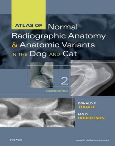
Atlas of Normal Radiographic Anatomy and Anatomic Variants in the Dog and Cat, 2 Edition
Atlas of Normal Radiographic Anatomy and Anatomic Variants in the Dog and Cat, 2 Edition. Becoming a proficient diagnostic radiologist is a long journey.
Atlas of Normal Radiographic Anatomy and Anatomic Variants in the Dog and Cat, 2 Edition

Specialty training leading to board certification entails at least 4 years of post-DVM structured learning, followed by a rigorous multistage examination. However, board-certified radiologists make up only a small fraction of all veterinarians who interpret radiographs each day. Most radiographic studies are interpreted by competent veterinarians whose training in image interpretation has been limited to relatively few contact hours of didactic instruction and supervised clinical training. All of these veterinarians, as well as students who are just beginning to develop their interpretive skills, must have a solid appreciation for normal radiographic anatomy, anatomic variants, and things that mimic disease, which are affectionately termed “fakeouts” by those of us who spend our lives interpreting images.
The vastness of normal variation within dogs and cats is staggering. Although the generic cat is relatively standard, dogs come in all shapes and sizes, with innumerable inherent variations that can be misinterpreted as disease unless recognized as normal. On top of this inherent varia-tion is the variation introduced by radiographic positioning that can lead to countless variations in the appearance of a normal structure. During their training, specialists have this information drilled into them during many hours of mentored learning and brow-beating by experienced radiologists. Nonspecialists, on the other hand, may have had some introduction to normal radiographic anatomy during veterinary school, but the acuity of recall becomes dulled by the sheer volume of memory-bank information needed to be a competent, licensed, contemporary veterinarian. During one’s education as a student, it is impossible to be exposed to the range of normal that is likely to be encountered in practice and then influenced by radiographic positioning. Therefore there is a real need for a reference source for practicing veterinarians and students to assist them in the daunting task of interpreting clinical radiographs competently. This need led to the development of this atlas.
In this book, we have not only pointed out the identity of essentially every clinically significant anatomic part of a dog or cat that can be seen radiographically, we have also included more than one example of those parts where normal inherent variation can confuse interpretation. Simply labeling structures in radiographs of a generic dog or cat is highly inadequate in addressing the mission of providing a clinically relevant resource. Additionally, this atlas includes context relevant to the description of normal anatomy that only a radiologist can provide. Normal is presented in the context of how it is modified by the procedure of making the radiograph. Although this is not a radiographic positioning guide, specific technical factors have been included to the extent that their influence on the image is so great that they must be understood for the image to be interpreted accurately.
Finally, this book is not simply a picture atlas. Every body part is put into context with a textual description. This provides a basis for the reader to understand why a structure appears as it does in radiographs, and it enables the reader to appreciate variations of normal that are not included based on an understanding of basic radiographic principles. This may require a bit of effort from the reader in comparison to a picture atlas, but this small investment of time has the potential for a big payoff in terms of interpretive ability.
[expand title=” “]
[/expand]
Password: pdflibrary.net


