Manual of Canine and Feline Musculoskeletal Imaging, The original Manual of Small Animal Diagnostic Imaging, edited by Professor Robin Lee, has for many years been an invaluable source of information for veterinary undergraduates as well as an important presence on the practice bookshelf.
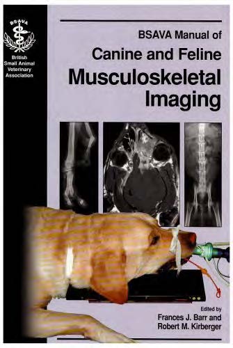
Manual of Canine and Feline Musculoskeletal Imaging
However, diagnostic imaging has rapidly developed and expanded over the last 10-20 years. Ultrasonography as an imaging modality has been available for some years, but as technology improves and the body of experience and knowledge grows, the range of potential applications widens. CT and MR are advanced cross-sectional imaging modalities which the general public are increasingly aware of, and thus we all need to be aware of the situations in which such modalities may be usefully applied. Despite the explosion in ‘other imaging modalities’, radiography remains as the day to day diagnostic imaging modality of choice in many practices.
It became clear to us that the original concept of the Manual of Small Animal Diagnostic Imaging should be retained -but expanded and improved upon. As the amount of knowledge is vast it could not all be accommodated into one manual, thus the concept of the Manual of Canine and Feline Musculoskeletal! Imaging was born to be followed in due course by its sister manuals of Thoracic Imaging and Abdominal-Imaging. Current small animal imaging textbooks tend to either include horse radiology or vast amounts of ultrasonography. These manuals will be ideal for those practitioners who do not have an interest in all the species or differing imaging modalities. It also prevents the practitioner having to buy a text that includes the thorax or abdomen if he/she rarely makes radiographs of these body regions.
This manual is extremely practical to use being structured along anatomical lines, with chapters considering each joint, the long bones and the skull and spine. There are additional general chapters considering the soft tissues, bone and joints which are common to all regions of the musculoskeletal system. We have concentrated on radiography and radiology. It is of vital importance that radiographs are of good technical quality so as to maximize the diagnostic information available.
So each chapter contains sections on radiographic technique, as well as normal radiographic anatomy, before considering the radiological features which may be associated with injury or disease. A vital part of this new Manual is the illustrations; we have kept part of the concept of the original manual with clear line drawings, but added a wide range of radiographs to complement the text. Although radiography and radiology are central, each chapter considers alternative imaging techniques and the situations in which they may be particularly useful, in order to guide the reader appropriately when further diagnostic information is needed.
Direct Link For Free Membership: –
| File Size: | 39 MB | |
| Download Link: | Click Here | |
| Password: | PDFLibrary.Net (if Required) | |